接第一篇:
Materials and methodology
Chemicals and microorganisms.
The EP standard was purchased from Yunnan Xuli Biological Technical Co., Ltd. Methanol of the chromatographic grade was obtained from Thermo Fisher Technology Co., Ltd. Other reagents were of analytical grade or reagent grade and provided by the Sinopharm Chemical Reagent Co., Ltd., Shanghai, China.
P. cicadae strain was purchased by the China Microbiological Culture Collection Center (NO. bio-33088), maintained on the new potato dextrose agar (PDA) slant, stored at 4 °C, and passaged once every 3 months.
Culture medium.
The seed medium was a PDA medium composed of (g/L) 20 glucose, 4 yeast powder, 3 peptone, 1 KH2PO4, and 1 MgSO4. The basal fermentation medium was composed of (g/L) 200 fresh potato, 40 glucose, 4 peptone, 1 KH2PO4, and 2 MgSO4. All media were sterilized at 121 °C for 30 min in an autoclave.
Inoculation and fermentation.
Erlenmeyer flasks (250 mL) containing 100 mL seed medium were inoc- ulated and incubated in a rotary shaker at 25 °C and 120 rpm for 72 h to prepare the inoculums. A 10% (v/v) inoculum was aseptically inoculated to 100 mL fermentation medium. The fermentation was carried out in 250- mL Erlenmeyer flasks in a rotary shaker at 25 °C and 120 rpm for 48 h. The cultured samples were centrifuged (6000×g for 15 min, 4 °C) to precipitate the biomass. The biomass was dried to constant weight at 80 °C for EP analysis.
Experiment design.
The mono‑factor at a time. Growth and EP production were studied with glucose, sucrose, maltose, fructose mannitol, or glycerol as a carbon source at a concentration of 20 g/L (with the other constituents same as those in the basal fermentation medium). Different nitrogen sources [peptone, urea, yeast extract, NH3·H2O, and (NH4)2SO4] were implemented to basal fermentation medium at a concentration of 3 g/L, The medium without nitrogen source was used as the control. The tested inorganic salts included (g/L) KH2PO4 (1), MgSO4 (1), (mg /L) MnSO4 (20), CuSO4 (20), ZnSO4 (20), and FeSO4 (20). The cultures were incubated in a rotary shaker at 25 °C and 120 rpm for 48 h. After 48 h, the flasks were harvested to analyze the dry cell weight and EP yield.
Uniform design.
Optimization studies were carried out based on a uniform design. Six independent variables were the concentrations of glycerol, yeast powder, peptone, ZnSO4, MgSO4, and KH2PO4. Sampling time was used as the control variable. Each independent variable was assessed at 10 different levels, resulting in a U10(10)6 (Table 1). All experiments were carried out in 250-mL Erlenmeyer flasks containing 100 mL medium. The cul- tures were carried out in a rotary shaker at 25 °C and 120 rpm for 48 h. Three flasks of each experiment were harvested at 28, 56, 84, and 112 h to analyze dry cell weight and EP yield.
Kinetic model.
Two types of models (such as structured and unstructured models) were applied to describe a microbial process. Compared with the unstructured model, a structured model reveals more infor- mation on physiological characterization, composition, and regulatory adaptations to the environment of the microorganisms45. The viscous fermentation broth model (Contois model), substrate inhibition model (Andrews model), and product inhibition model (Aibe model) are a partial link between cell growth and viscosity of fer- mentation broth, substrate concentration, or product concentration, respectively. These models were used in our work.
Model assumptions.
Kinetic models of microbial growth were carried out based on the Monod model, Andrews model, Contois model, and Aibe model. To describe the effect of glycerol concentration on biomass growth, the following considerations were assumed46.
(1) The mycelium ball is thought to be a tiny chemical reactor. In the reactor, the substrate is converted into mycelia by a complex network of enzyme-catalyzed reactions. In this process, there is no mass transfer resistance inside the mycelium ball.
(2) In the complex network of enzyme-catalyzed reactions, glycerol is the only limiting substrate. In all meta- bolic pathways converting glycerol into mycelia and metabolite, only one pathway is the limiting pathway, in which the velocity is the slowest. In all reactions of the limiting pathway, only one key step reaction controls the whole velocity of the limiting pathway. Namely, the biomass growth velocity depends on the enzyme reaction of the key step and the effect of the glycerol concentration on the limiting reaction rate.
(3) The enzyme concentration of the limiting reaction is proportional to the concentration of the mycelium ball, and the concentration of the mycelium ball is proportional to the consumption concentration of the limiting substrate. The growth velocity of the mycelium ball is proportional to the consumption velocity of the limiting substrate, and the concentration of the product is proportional to the consumption concentra- tion of the limiting substrate.
Based on these assumptions, the Monod model, Andrews model (inhibition model by substrate), Contois model (inhibition model by viscosity), and Aibe model (inhibition model by production) can be used to fit the biomass data in the progress of P. cicadae.
Monod model, Contois model, Andrews model, and Aibe model were expressed by the following equations, respectively:
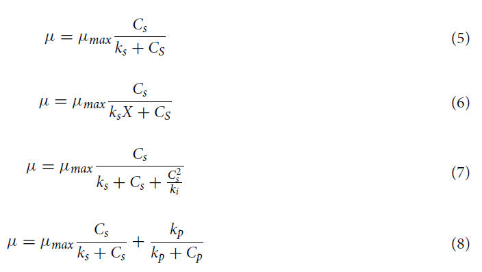
In these models, μ and μmax are the specific growth rate and the maximum specific growth rate (h−1), respec- tively; Cs is the substrate concentration, namely glycerol concentration; ks is the saturation constant (the substrate concentration at the half of the maximum specific growth rate, mM), X is the biomass concentration, ki and kp are the inhibition constants of substrate and inhibition product (mM), respectively, and Cp is the concentration of the inhibition product.
According to the definition of specific growth rate, μ can be expressed by the following equation:

where dX/dt is the biomass growth rate.
According to the above-mentioned assumptions, there are the following equations:


where Yb and Yp are the biomass and product yield coefficients on the substrate (glycerol), respectively.
After substituting Eqs. (5)–(8) in Eqs. (1)–(4), followed by integration and rearrangement, the following new Monod model (Eq. 9), Contois model (Eq. 10), Andrews model (Eq. 11), and Aibe model (Eq. 12) were given.
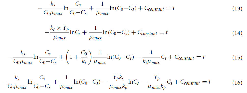
These equations were applied to fit the data of kinetic experiments.
The model solution and simulations.
Experimental data from batch fermentation in a 20-L mechanically stirred fermentor were utilized to simulate the kinetic parameters by the developed model Eqs. (9)–(12). The fermenta- tion medium was the optimized medium. The fermentation conditions of the 20-L mechanically stirred fermen- tor were set as follows: medium volume 15 L, inoculum 10%, rotational speed 120 rpm, aeration rate 15 L/min, and temperature 25 °C.
Verification of kinetic inference.
The verification tests were carried in a 20-L mechanically stirred fermentor. The three experiments were designed in agreement with the kinetic experiments. Water and glycerol were not supplied in the group 1 experiment, and 1 L water was supplied at 40 h and 60 h in the process of fermentation in the group 2 experiment. In the group 3 experiment, 900 g glycerol was added in three batches, 300 g was added to the medium before sterilization, and another 600 g glycerol (2,000 mL 30% glycerol aqueous solution) was added at 40 h and 60 h, respectively. The fermentation conditions were the same as the kinetic conditions and fermentation time 80 h.
Analytical methods.
Preparation of sample solution. Briefly, the biomass of P. cicadae was dried to con- stant weight and powdered in a grinding mill. Next, 0.10 g biomass powder was loaded into a 5-mL centrifuge tube and mixed with 3 mL MeOH. The tube was sonically extracted at 25 °C for 30 min. The conditions of ultra- sonic treatment were set as follows: frequency 40 Hz and power 50 W. The tube was centrifuged at 1.3 × 10⁴ rpm for 2 min. The supernatant was subjected to LC–MS/MS analysis. The extraction was performed in triplicate.
Preparation of standard EP stock solution (50 μg/mL).
Briefly, 10 mg standard EP was dissolved in 200 mL methanol to prepare a standard EP stock solution. Before use, the stock solution was diluted with methanol to prepare the working solutions of various concentrations.
Chromatographic conditions of LC–MS/MS.
An HPLC–MS/MS (Agilent 1290 UPLC/6540 Q-Tof, USA) system equipped with an Acquity UPLC BEH C18 column (130 Å, 1.7 μm, 2.1 × 100 mm) was used in the present deter- mination. A gradient elution procedure was applied to the elution of EP. The mobile solvent A and solvent B used in the gradient elution procedure were 0.1% formic acid–methanol and 0.1% formic acid–water, respectively. The elution procedure was programmed as follows: 10% A to 95% A (0–2.5 min), 95% A to 95% A (2.5–10 min), 95% A to 10% A (10–10.5 min), and 10% A to 10% A (10.5–12.5 min). Other conditions were set as follows: flow rate 0.2 mL/min, column temperature 40 °C, injection volume 5 μL, and sample temperature 20 °C. The quantization was operated in the multi-response monitoring (MRM) mode with a positive ion mode to monitor the precursor-production ion pair transitions of m/z 429.6–393.6 for EP.
Data analysis.
Non-linear multiple regression analyses have been utilized in the optimization of medium components and the parameters of reaction process to find the optimum medium and to determine the opti- mum reaction process, which often seem to provide higher accuracy47,48. The p-value, F-value and R2 derived from Statistical Computing software were used to apply multi-variable non-linearregression analysis. In the study, after a series of modeling, according tot p-value, F-value and R2, the optimum medium and reaction process were found.
One way analysis of variance (Anova) is a method to analyze the results of univariate analysis and to test whether test factors have a significant effect on test results. One-way ANOVA has proven its effectiveness in solving the problem of high dimensionality in the feature space49. Thus, the one way Anova was applied into the study as a filter method to select the relevant features.
All data were expressed as means ± standard errors (in the mono-factor at time and the Uniform-design experimentation) or as means (in the kinetic model experiment). All the multi-ple non-linear regression equa- tions were conducted using SPSS 17.0 software (IBM, Ammonst City, USA) and Microsoft Excel (Microsoft, Redmond City, USA), the analysis of variance (ANOVA) was performed by Dunnett’s test, and p < 0.05 was considered as statistically significant.
Conclusions
The optimal medium for maximum production of EP by P. cicadae was determined by a combination of a mono-factor experiment, a uniform design method, and a non-linear regression. This was the first report on EP production by P. cicadae fermentation. The optimum fermentation time was 80 h. The maximum flask culture yield of EP yield (256 μg/L) after optimization was increased by nearly five-fold compared with that before the optimization, which was also higher than the maximum theoretical EP yield (203.92 μg/L). The combinational use of four structured models indicated that glycerol and water could further increase the yield of EP in the fermentation process of P. cicadae.
Received: 29 August 2021; Accepted: 31 January 2022
Published online: 07 April 2022
References
1. Kim, S., Park, S., Min, T. & Yu, K. Antioxidant activity of ergosterol peroxide (5,8-epidioxy-5α, 8α-ergosta-6, 22E-dien-3-ol) in
Armillariella mellea. Bull. Korean Chem. Soc. 20, 819–823 (1999).
2. Kobori, M., Yoshida, M., Ohnishi-Kameyama, M. & Shinmoto, H. Ergosterol peroxide from an edible mushroom suppresses inflammatory responses in RAW264.7 macrophages and growth of HT29 colon adenocarcinoma cells. Br. J. Pharmacol. 150, 209–219 (2007).
3. Yasukawa, K. et al. Inhibitory effects of ergosterol isolated from the edible mushroom Hypsizigus marmoreus on TPA-induced inflammatory ear oedema and tumour promotion in mice. Phytother. Res. 8, 10–13 (1994).
4. Fujimoto, H., Nakayama, M., Nakayama, Y. & Yamazaki, M. Isolation and characterization of immunosuppressive components of three mushrooms, Pisolithus tinctorius, Microporus flabelliformis and Lenzites betulina. Chem. Pharm. Bull. (Tokyo) 42, 694–697 (1994).
5. Kuo, Y. C., Weng, S. C., Chou, C. J., Chang, T. T. & Tsai, W. J. Activation and prolifer-ation signals in primary human T lymphocytes inhibited by ergosterol peroxideisolated from Cordyceps cicadae. Br. J. Pharmacol. 140, 895–906 (2003).
6. Takei, T., Yoshida, M., Ohnishi-Kameyama, M. & Kobori, M. Ergosterol peroxide, an apoptosis-inducing component isolated from Sarcodon aspratus (Berk.) S. Ito. Biosci. Biotechnol. Biochem. 69, 212–215 (2005).
7. Wu, Q. P. et al. Ergosterol peroxide isolated from Ganoderma lucidum abolishes microRNA miR-378-mediated tumor cells on chemoresistance. PLoS ONE 7, e44579 (2012).
8. Zhu, R., Zheng, R., Deng, Y. Y., Chen, Y. & Zhang, S. Ergosterol peroxide from Cordyceps cicadae ameliorates TGF- 1-induced activation of kidney fibroblasts. Phytomedicine 21, 372–378 (2014).
9. Kang, J. H. et al. Ergosterol peroxide from Chaga mushroom (Inonotus obliquus) exhibits anti-cancer activity by down-regulation of the β-catenin pathway in colorectal cancer. J. Ethnopharmacol. 173, 303–312 (2015).
10. Krzyczkowski, W. et al. Isolation and quantitative determination of ergosterol peroxide in various edible mushroom species. Food Chem. 113, 351–355 (2009).
11. Paterson, R. R. Cordyceps: a traditional Chinese medicine and another fungaltherapeutic biofactory. Phytochemistry 69, 1469–1495 (2008).
12. Wojcikowski, K., Johnson, D. W. & Gobe, G. Medicinal herbal extracts—renalfriend or foe? Part two: Herbal extracts with potential renal benefits. Nephrology (Carlton) 9, 400–405 (2004).
13. Wojcikowski, K., Johnson, D. W. & Gobe, G. Herbs or natural substances as com-plementary therapies for chronic kidney disease: Ideas for future studies. J. LabClin. Med. 147, 160–166 (2006).
14. Jin, Z. & Chen, Y. Clinical observation on Cordyceps Cicadae Shing Tang in pre-venting the progression of chronic renal failure. Chin. Arch. Tradi. Chin. Med. 24, 1457–1459 (2006).
15. Zhou, H. J., Yi, P. C. & Yue, Y. D. The mechanism study of Cordyceps sobolif-era mycelium preventing the progression of glomeru- losclerosis. Chin. J. Integr. Tradit. Western Nephrol. 6, 132–136 (2005).
16. Zhu, R. et al. Cordyceps cicadae extracts ameliorate renal malfunction in a remnant kidney model. J. Zhejiang Univ. Sci. B 12, 1024–1033 (2011).
17. He, L., Shi, W., Liu, X., Zhao, X. & Zhang, Z. Anticancer action and mechanism of ergosterol peroxide from Paecilomyces cicadae fermentation broth. Int. J. Mol. Sci. 19, 3935 (2018).
18. Betancourt, M. A. M., Santamaria, C. A. L., Gómez, M. L. & Caranton, A. R. G. Experimental analysis of biodiesel synthesis from palm kernel oil: Empirical model and surface response variables. React. Kinet. Mech. Cat. 131, 297–317 (2020).
19. Dai, C. & Lei, X. J. An improvement decomposition-based multi-objective evolutionary algorithm with uniform design. Knowl‑ Based Syst. 125, 108–115 (2017).
20. González-Vega, R. I. et al. Optimization of growing conditions for pigments production from microalga Navicula incerta using response surface methodology and its antioxidant capacity. Saudi J. Biol. Sci. 28, 1401–1416 (2021).
21. Kumar, M. V., Kumar, B. J. K. & Rudresha, N. Optimization of machining parameters in CNC turning of stainless steel (EN19) by Taguchi’s orthogonal array experiments. Mater. Today: Proc. 5, 11395–11407 (2018).
22. Xiong, Q., Li, Z., Luo, H. & Zhao, Z. Wind tunnel test study on wind load coefficients variation law of heliostat based on uniform design method. Sol. Energy 184, 209–229 (2019).
23. Zhou, S. K., Bi, T. N., Xu, Y. F., Zhang, R. L. & Yang, M. J. Extraction optimization of carbohydrate compound from Huangqi using orthogonal design. Int. J. Biol. Macromol. 58, 13–17 (2013).
24. Fang, K. T., Lin, D. K. J., Winker, P. & Zhang, Y. Uniform design: Theory and application. Technometrics 42, 237–248 (2002).
25. Tan, Y. Y., Jiao, Y. C., Li, H. & Wang, X. K. MOEA/D + uniform design: A new version of MOEA/D for optimization problems with many objectives. Comput. Oper. Res. 40, 1648–1660 (2013).
26. Ahmad, F., Jameel, A. T., Kamarudin, M. H. & Mel, M. Study of growth kinetic & modeling of ethanol production by Saccharomyces cerevisiae. Afr. J. Biotechnol. 16, 18842–18846 (2011).
27. Lei, F., Rotbøll, M. & Jørgensen, S. B. A biochemically structured model for Saccharomyces cerevisiae. J. Biotechnol. 88, 205–221 (2001).
28. Liu, C. G., Lin, Y. H. & Bai, F. W. A kinetic growth model for Saccharomyces cerevisiae grown under redox potential-controlled very-high-gravity environment. Biochem. Eng. J. 56, 63–68 (2011).
29. Sansonetti, S., Hobley, T. J., Calabrὸ, V., Villadsen, J. & Sin, G. A biochemically structured model for ethanol fermentation by Kluveromyces marxianus: A batch fermentation and kinetic study. Bioresour. Technol. 102, 7513–7520 (2011).
30. Surendhiran, D., Vijay, M., Sivaprakash, B. & Sirajunnisa, A. Kinetic modeling of microalgal growth and lipid synthesis for biodiesel production. 3 Biotechnol 5, 663–669 (2015).
31. Sharma, V. & Mishra, H. N. Unstructured kinetic modeling of growth and lactic acid production by Lactobacillus plantarum NCDC 414 during fermentation of vegetable juices. LWT Food Sci. Technol. 59, 1123–1128 (2014).
32. Liu, X. Z. & Chen, S. Y. Nutritional requirements of the nema-tophagous fungus Hirsutella rhossiliensis. Biocontrol Sci. Technol. 12, 381–393 (2002).
33. Clotilde, T. et al. Minimal information for studies of extracellular vesicles 2018 (MISEV2018): A position statement of the Inter- national Society for Extracellular Vesicles and update of the MISEV2014 guidelines. J Extracel. vesicles 7, 1535750 (2018).
34. MacCabe, A. P., Ninou, E. I., Pardo, E. & Orejas, M. Catabolism of L-rhamnose in A. nidulans proceeds via the non-phosphorylated pathway and is glucose repressed by a CreA-independent mechanism. Microb. Cell Fact. 19, 188 (2020).
35. Yang, M., Ye, J., Qin, H., Long, Y. & Li, Y. Influence of perfluorooctanoic acid on proteomic expression and cell membrane fatty acid of Escherichia coli. Environ. Pollut. 220, 532–539 (2017).
36. Liu, H. et al. Influence analysis of glycerol in fumaric acid co-fermentation process by Rhizopus arrhizus. J. Environ. Chem. Eng.9, 104750 (2021).
37. Quartacci, M. F., Glisic, O., Steranovic, B. & Navarri-Izzo, F. Plasma membrane lipids in the resurrection plant Ramonda serbica following dehydratation. J. Exp. Bot. 53, 2159–2166 (2002).
38. Xu, F., Tao, W. Y., Cheng, L. & Guo, L. Strain improvement and optimization of themedia of taxol-producing fungus Fusarium maire. Biochem. Eng. J. 31, 67–73 (2006).
39. Caddick, M. X. Nitrogen regulation in mycelial fungi. In Biochemistry and Molecular Biology (eds Brambl, R. & Marzluf, G. A.) 349–368 (Springer, 2004).
40. Ehrlich, K. C. & Cotty, P. J. Variability in nitrogen regulation of aflatoxin production by Aspergillus flavus strains. Appl. Microbiol. Biotechnol. 60, 174–178 (2002).
41. Morschhäuser, J. Nitrogen regulation of morphogenesis and protease secretion in Candida albicans. In. J. Med. Microbiol. 301, 390–394 (2011).
42. Xiao, J. H. et al. Enhanced simultaneous production of mycelia and intracellular polysaccharide in submerged cultivation of Cordyceps jiangxiensis using desirability functions. Proc. Biochem. 41, 1887–1893 (2006).
43. Feng, J. et al. An unstructured kinetic model for the improvement of triterpenes production by Ganoderma lucidum G0119 based on nitrogen source effect. Biotechnol. Bioproc. E 19, 727–732 (2014).
44. Tian, X. et al. Kinetic analysis of sodium gluconate production by Aspergillus niger with different inlet oxygen concentrations. Bioproc. Biosyst. Eng. 41, 1697–1706 (2018).
45. Ochoa, S., Yoo, A. & J. U., Wozny, G., & Yang, D. R. M.,. Modeling and parameter identification of the simultaneous saccharifica- tion -fermentation process for ethanol production. Biotechnol. Prog. 23, 1454–1462 (2007).
46. Stanbury, P. F., Whitaker, A. & Hall, S. J. Microbial Growth Kinetics (Principles of Fermentation Technology (Third Edition) Aca- demic press, 2016).
47. Gomathi Devi, L. & EraiahRajashekhar, K. A kinetic model based on non-linear regression analysis is proposed for the degradation of phenol under UV/solar light using nitrogen doped TiO2. J. Mol. Catal. A Chem. 334, 65–76 (2011).
48. Zhang, Z. et al. The optimum conditions and mechanism for increasing exo-polysaccharide production of Truffles melanosporum by Dioscorea saponins. LWT Food Sci. Technol. 107, 331–339 (2019).
49. Elssied, N. O. F., Ibrahim, O. & Osman, A. H. A novel feature selection based on one-way ANOVA F-test for e-mail spam clas- sification. Res. J. Appl. Sci. Eng. Technol. 7, 625–638 (2014).
Acknowledgements
This work was financially supported by the China Postdoctoral Science Foundation (No. 2015M571691), and the National Natural Science Foundation of China (No. 31301544).
Author contributions
Qian J and Zhang Z designed the whole experiment; Shi F, Qian J and Zhang Z wrote the mainmanuscript text; He L, Shi F and Zheng H did the whole tests; Zheng H prepared the whole figure; Shi H prepared the whole table. Zhang Z provided the fund of the whole study.
Competing interests
The authors declare no competing interests.
Additional information
Correspondence and requests for materials should be addressed to J.Q. or Z.Z.
Reprints and permissions information is available at www.nature.com/reprints.
Publisher’s note Springer Nature remains neutral with regard to jurisdictional claims in published maps and institutional affiliations.
Open Access
This article is licensed under a Creative Commons Attribution 4.0 International License, which permits use, sharing, adaptation, distribution and reproduction in any medium or format, as long as you give appropriate credit to the original author(s) and the source, provide a link to the Creative Commons licence, and indicate if changes were made. The images or other third party material in this article are included in the article’s Creative Commons licence, unless indicated otherwise in a credit line to the material. If material is not included in the article’s Creative Commons licence and your intended use is not permitted by statutory regulation or exceeds the permitted use, you will need to obtain permission directly from the copyright holder. To view a copy of this licence, visit http://creativecommons.org/licenses/by/4.0/.
© The Author(s) 2022
Terms and Conditions
Springer Nature journal content, brought to you courtesy of Springer Nature Customer Service Center GmbH (“Springer Nature”).
Springer Nature supports a reasonable amount of sharing of research papers by authors, subscribers and authorised users (“Users”), for small- scale personal, non-commercial use provided that all copyright, trade and service marks and other proprietary notices are maintained. By accessing, sharing, receiving or otherwise using the Springer Nature journal content you agree to these terms of use (“Terms”). For these purposes, Springer Nature considers academic use (by researchers and students) to be non-commercial.
These Terms are supplementary and will apply in addition to any applicable website terms and conditions, a relevant site licence or a personal subscription. These Terms will prevail over any conflict or ambiguity with regards to the relevant terms, a site licence or a personal subscription (to the extent of the conflict or ambiguity only). For Creative Commons-licensed articles, the terms of the Creative Commons license used will apply.
We collect and use personal data to provide access to the Springer Nature journal content. We may also use these personal data internally within ResearchGate and Springer Nature and as agreed share it, in an anonymised way, for purposes of tracking, analysis and reporting. We will not otherwise disclose your personal data outside the ResearchGate or the Springer Nature group of companies unless we have your permission as detailed in the Privacy Policy.
While Users may use the Springer Nature journal content for small scale, personal non-commercial use, it is important to note that Users may not:
1. use such content for the purpose of providing other users with access on a regular or large scale basis or as a means to circumvent access control;
2. use such content where to do so would be considered a criminal or statutory offence in any jurisdiction, or gives rise to civil liability, or is otherwise unlawful;
3. falsely or misleadingly imply or suggest endorsement, approval , sponsorship, or association unless explicitly agreed to by Springer Nature in writing;
4. use bots or other automated methods to access the content or redirect messages
5. override any security feature or exclusionary protocol; or
6. share the content in order to create substitute for Springer Nature products or services or a systematic database of Springer Nature journal content.
In line with the restriction against commercial use, Springer Nature does not permit the creation of a product or service that creates revenue, royalties, rent or income from our content or its inclusion as part of a paid for service or for other commercial gain. Springer Nature journal content cannot be used for inter-library loans and librarians may not upload Springer Nature journal content on a large scale into their, or any other, institutional repository.
These terms of use are reviewed regularly and may be amended at any time. Springer Nature is not obligated to publish any information or content on this website and may remove it or features or functionality at our sole discretion, at any time with or without notice. Springer Nature may revoke this licence to you at any time and remove access to any copies of the Springer Nature journal content which have been saved.
To the fullest extent permitted by law, Springer Nature makes no warranties, representations or guarantees to Users, either express or implied with respect to the Springer nature journal content and all parties disclaim and waive any implied warranties or warranties imposed by law, including merchantability or fitness for any particular purpose.
Please note that these rights do not automatically extend to content, data or other material published by Springer Nature that may be licensed from third parties.
If you would like to use or distribute our Springer Nature journal content to a wider audience or on a regular basis or in any other manner not expressly permitted by these Terms, please contact Springer Nature at onlineservice@springernature.com