Complete genome sequence of a novel victorivirus infecting cicada flower
(Cordyceps chanhua)
Qiuyan Zhu1 · Najie Shi1 · Yuxiang Zhang1 · Fan Peng1 · Guogen Yang2 · Bo Huang1
Received: 11 June 2022 / Accepted: 25 August 2022 / Published online: 20 December 2022
© The Author(s), under exclusive licence to Springer-Verlag GmbH Austria, part of Springer Nature 2022
Abstract
Cicada flower, Cordyceps chanhua, is a precious edible and medicinal mushroom with uses in both medicine and food in China. In this study, Cordyceps chanhua strain RCEF5995 was found to be coinfected by a previously characterized alternavirus, Cordyceps chanhua alternavirus 1 (CcAV1), and a novel victorivirus, tentatively named "Cordyceps chanhua victorivirus 1" (CcV1). Molecular characterization of CcV1 showed that its complete genome is 5,232 nucleotides long with a GC content of 57.5%. Sequence analysis indicated that CcV1 contains two overlapping open reading frames (ORFs), ORF1 and ORF2, encoding a putative coat protein (CP) of 742 amino acids (aa) and a putative RNA-dependent RNA polymerase (RdRp) of 836 aa, respectively. The termination codon of the CP ORF overlaps with the initiation codon of the RdRp ORF at the tetranucleotide sequence AUGA. Homolog searches, sequence comparisons, and phylogenetic analysis based on deduced amino acid sequences of RdRp indicated that CcV1 is a new member of the genus Victorivirus, family Totiviridae.
Handling Editor: Stephen John Wylie.
Qiuyan Zhu and Najie Shi contributed equally to this study and share first authorship.
E-mail Bo Huang:bhuang@ahau.edu.cn
1 Anhui Provincial Key Laboratory of Microbial Pest Control, Anhui Agricultural University, Hefei 230036, China
2 School of Plant Protection, Anhui Agricultural University, Hefei 230036, China
Cicada flower, known as an edible and medicinal mushroom, is the fruiting body of the soil-dwelling fungus Cordyceps chanhua (syn. Isaria cicadae and Paecilomyces cicadae), which is naturally parasitic on cicada nymphs of several species in the family Cicadidae [1, 2]. This fungus–host insect complex, as a noted traditional Chinese medicine, has long been applied in relieving exhaustion and treating numerous diseases [1]. Moreover, a modern pharmacological study has shown that many bioactive compounds isolated from cicada flower have antitumor, antifatigue, anti-inflammatory, immunomodulatory, analgesic, renoprotective, and sedative properties [3]. To meet the great consumer demand for cicada flower, large-scale cultivation of coremium of Cordyceps chanhua has been established in China [4].
Mycoviruses are widespread among all fungal groups, with the majority having a double-stranded RNA (dsRNA) genome. Taxonomically, dsRNA mycoviruses have been assigned to the families Amalgaviridae, Chrysoviridae, Megabirnaviridae, Partitiviridae, Polymycoviridae, Quadriviridae, Spinareoviridae, and Totiviridae and the genus Botybirnavirus (https:// ictv. global/ taxon omy). Currently, the International Committee on Taxonomy of Viruses (ICTV) recognizes five genera in the family Totiviridae: Totivirus, Victorivirus, Giardiavirus, Trichomonasvirus, and Leishmaniavirus [5, 6]. Victoriviruses, which primarily infect fungi, possess a single linear dsRNA genome 4.6-6.7 kb in length, containing two overlapping ORFs encoding a capsid protein (CP) and an RNA-dependent RNA polymerase (RdRp) [7, 8]. Its dsRNA is encapsidated in a non-enveloped, icosahedral virion approximately 30-40 nm in diameter [9].
Fungal viruses that infect Cordyceps chanhua have not been studied extensively. So far, only two dsRNA viruses have been isolated from C. chanhua, an alternavirus and an unassigned partitivirus [10]. In this study, we demonstrated that C. chanhua was coinfected by a previously characterized alternavirus, Cordyceps chanhua alternavirus 1 (CcAV1, 100% aa sequence identity in the putative RdRp) and a novel dsRNA virus belonging to the genus Victorivirus, which we have designated as "Cordyceps chanhua victorivirus 1" (CcV1).
Entomopathogenic fungi were collected in the Guniujiang National Forest Park in Anhui Province, China, in July 2021. Strain RCEF5995, isolated from cicada, was identified as C. chanhua through combined morphological and ITS sequence data (GenBank accession number OP271288). The fungus was cultured on SDAY medium (1% w/v peptone, 4% w/v dextrose, 0.2% w/v yeast, and 1.5% w/v agar) at 25 °C for 5 days for dsRNA multiplication [11], and dsRNA was extracted using CF-11 cellulose (Sigma) chromatography and purified by S1 nuclease and DNase I treatment (TaKaRa, Dalian, China) as described previously [12]. The purity and size of the dsRNA were determined by electrophoresis on a 1.0% agarose gel, which indicated that strain RCEF5995 contains four distinct dsRNAs, approximately 5.2, 3.5, 2.6, and 2.4 kb in length, named dsRNA 1-4, respectively (Fig. 1A). To further analyze these dsRNAs, we performed high-throughput sequencing of all gel-purifed dsRNAs on an Illumina HiSeq 2500 platform at BGI (Shenzhen, China). The clean reads were assembled into approximately 4,323 contigs with an N50 value of 1,123, using Trinity v2.1.1, and BLAST analysis was conducted using viral reference sequences from the NCBI database. BLAST and RT-PCR results confirmed that strain RCEF5995 is coinfected by two different dsRNA mycoviruses: a victorivirus (dsRNA1) and the previously characterized alternavirus CcAV1 (dsRNA 2, dsRNA 3, and dsRNA 4). The primers for amplification of dsRNA1 and dsRNA2 are listed in Supplementary data S1.
To complete the 5′- and 3′-terminal genome sequences, amplification of the 5′ and 3′ cDNA ends was performed using RNA-ligase-mediated rapid amplification of cDNA ends (RLM-RACE) from a cDNA library constructed using two specific primers [13]. At least three PCR products cloned into the vector pMD18-T (Takara Bio Inc., Kusatu, Shiga) were sent to Sangon Biotech for Sanger sequencing to verify the sequences. The complete genome sequence of CcV1 was deposited in the GenBank database under the accession number ON804485. The GC content was determined using OligoCalc [14]. Putative ORFs in the CcV1 genome were identified using ORF Finder at NCBI (https:// www. ncbi. nlm. nih. gov/ orfnd er). Mfold was used to find potential secondary structures in the terminal sequences of the CcV1 genome [15]. The predicted RdRp amino acid sequence of CcV1 was aligned with those of other members of the genus Victorivirus, using the multiple sequence alignment program MAFFT [16]. A maximum-likelihood phylogenetic tree was constructed using MEGA X with the LG+G+I+F model and 1000 bootstrap replicates [17].
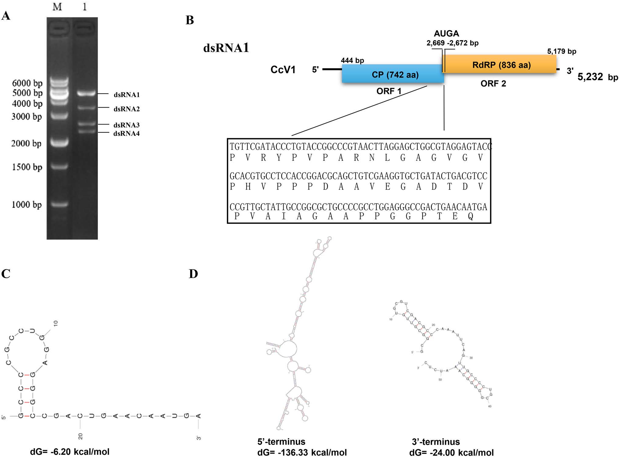
Fig. 1 Analysis of the dsRNA profile and genomic organization of the novel mycovirus (CcV1) from Cordyceps chanhua strain RCEF5995. (A) Purified dsRNA extracted from RCEF5995 was electrophoresed in a 1.5% agarose gel. M, DNA marker (10,000-bp ladder, Dongsheng Biotech); lane 1, dsRNAs. (B) Schematic representation of CcV1, with the amino acid sequence of the C-terminal region of ORF1 showing the Ala/Gly/Pro-rich stretch. (C) Schematic representation of the predicted H-type RNA pseudoknot 30 nt directly upstream of the AUGA motif (underlined) in CcV1. (D) Stable secondary structure of the 5’-terminus and 3’-terminus of CcV1.
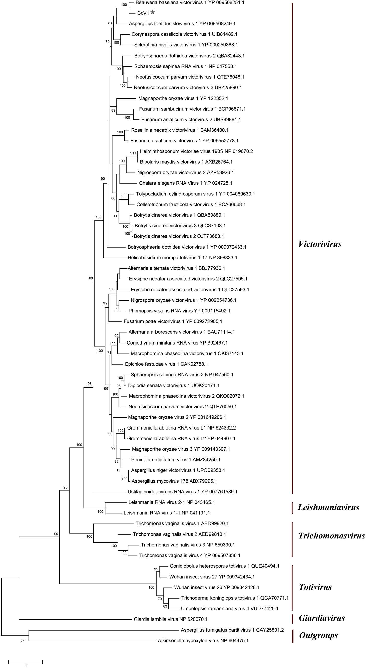
Fig. 2 Phylogenetic tree based on RdRp sequences from members of the family Totiviridae, constructed by the maximumlikelihood (ML) method, using the LG+G+I+F amino acid substitution model. The scale bar represents 1.0 amino acid substitutions per site, and numbers at the nodes indicate bootstrap support over 50% (1000 replicates).
The complete genome of CcV1 in strain RCEF5995 is 5,232 bp long with a predicted GC content of 57.5%, similar to other victorivirus genomes [18]. An ORF search revealed that the CcV1 genome is bicistronic. ORF1 is 2229 nt long (nt 444-2672), encoding a 742-amino-acid (aa) protein with a molecular weight of 78.39 kDa, while ORF2 is 2511 nt long (nt 2669-5179), encoding an 836-aa protein with a molecular weight of 91.09 kDa (Fig. 1B). Furthermore, the termination codon of ORF1 overlaps with the initiation codon of ORF2 at an AUGA tetranucleotide sequence at nt 2669-2672. This motif is used by many victoriviruses for translation of the downstream ORF2 by a termination-reinitiation mechanism [8]. An H-type pseudoknot structure was identified upstream of the AUGA motif in ORF1 (Fig. 1C). The untranslated regions (UTRs) at the 5' and 3' ends are 443 and 53 bp long, respectively, and were predicted to have stable secondary structures (Fig. 1D).
BLASTp analysis of the ORF1 and ORF2 protein showed significant similarity to victorivirus coat proteins (pfam05518) and RdRp proteins (RdRp_4, pfam02123). Furthermore, we found that the putative RdRp and CP of CcV1 had the highest sequence similarity to the corresponding proteins of Beauveria bassiana victorivirus 1 (70.74% and 73.42%, respectively). They also showed considerable sequence similarity to the CPs and RdRps of Ustilaginoidea virens RNA virus 1 (61.90%, 51.76%) and Aspergillus foetidus slow virus 1 (57.47%, 48.63%), suggesting that CcV1 is closely related to dsRNA mycoviruses in the genus Victorivirus, family Totiviridae. We also found an Ala/Gly/Pro-rich stretch in the C-terminal sequence of CcV1 CP (Fig. 1B), which is a characteristic feature of victoriviruses.
To determine the taxonomic status of CcV1, a maximumlikelihood (ML) phylogenetic tree based on RdRp amino acid sequences of 47 mycoviruses, including all members of the genus Victorivirus and representative members of the remaining four genera of the family Totiviridae, was constructed with two partitiviruses as outgroups. The RdRp-based phylogenetic tree demonstrated that five wellsupported clades corresponded to the established genera of the family Totiviridae, with high bootstrap values. Moreover, CcV1 formed a branch with members of the genus Victorivirus and was most closely related to and formed a distinct group with Beauveria bassiana victorivirus 1 (Fig. 2).
The cutoff values recommended by the ICTV for species demarcation within the genus Victorivirus are 60% pairwise identity in the RdRp and CP sequences. The RdRP and CP of CcV1 share 70.74% and 73.42% identity with the related proteins of Beauveria bassiana victorivirus 1, which is above this threshold. However, because CcV1 and Beauveria bassiana victorivirus 1 were isolated from different fungal host, we propose that CcV1 should nevertheless be considered a new member of the genus Victorivirus in the family Totiviridae based on phylogenetic analysis, RdRp sequence comparisons, and analysis of its termination-reinitiation strategy.
Supplementary Information The online version contains supplementary material available at https:// doi. org/ 10. 1007/ s00705- 022- 05640-2.
Funding This work was supported by the National Natural Science Foundation of China (Grant no. 32172473) and the Anhui Natural Science Foundation (Grant no. 1908085MC56).
Declarations
Conflict of interest The authors have no conflict of interest.
Ethical approval This article does not contain any studies with human participants or animals performed by any of the authors.
References
1. Li ZZ, Chen ZA, Chen YP (2014) Chanhua, a National Treasure cordyceps in China. Hefei University of Technology Press, Hefei
2. Fan WW, Zgang S, Zhang YJ (2019) The complete mitochondrial genome of the Chan-hua fungus Isaria cicadae: a tale of intron evolution in Cordycipitace. Environ Microbiol 21(2):864–879
3. Chunyu Y, Lu Z, Luo Z, Li S, Li H, Geng Y, Xu Hong XuZ, Shi J (2019) Promotion of metabolite synthesis in Isaria cicadae, a dominant species in the cicada flower microbiota, by Cicada Pupae. J Agric Food Chem 67:8476–8484
4. Dong C, Li W, Li Z, Yan W, Li T, Liu X (2016) Cordyceps industry in China: current status, challenges and perspectives-Jinhu declaration for cordyceps industry development. J Mycosystema 35:1–15
5. Ghabrial SA, Caston JR, Jiang D, Nibert ML, Suzuki N (2015) 50-plus years of fungal viruses. Virology 479–480:356–368
6. King AM, Adams MJ, Lefkowitz EJ et al (eds) (2011) Virus taxonomy. In: IXth report of the International Committee on Taxonomy of Viruses, 9th edn. Elsevier, Waltham
7. Jiang Y, Zhang T, Luo C, Jiang D, Li G, Li Q, Hsiang T, Huang J (2015) Prevalence and diversity of mycoviruses infecting the plant pathogen Ustilaginoidea virens. Virus Res 195:47–56
8. Li H, Havens WM, Nibert ML, Ghabrial SA (2011) RNA sequence determinants of a coupled termination-reinitiation strategy for downstream open reading frame translation in Helminthosporium victoriae virus 190S and other victoriviruses (Family Totiviridae). J Virol 85:7343–7352
9. Wickner RB, Ghabrial SA, Nibert ML, Patterson JL, Wang CC (2011) Family Totiviridae. In: King AMQ, Adams MJ, Carstens EB, Lefkowits EJ (eds) Virus taxonomy: ninth report of the international committee for the taxonomy of viruses. Elsevier, Academic Press, New York, pp 639–650
10. Zhang Y, Shi N, Wang P, Zhu Q, Yang G, Huang B (2022) Molecular characterization of a novel alternavirus infecting the entomopathogenic fungus Cordyceps chanhua. Arch Virol 167:1467–1470
11. Shi N, Yang G, Wang P, Wang Y, Yu D, Huang B (2019) Complete genome sequence of a novel partitivirus from the entomogenous fungus Beauveria bassiana in China. Arch Virol 164:3141–3144
12. Herrero N, Duenas E, Quesada-Moraga E, Zabalgogeazcoa I (2012) Prevalence and diversity of viruses in the entomopathogenic fungus Beauveria bassiana. Appl Environ Microbiol 78:8523–8530
13. Coutts RHA, Livieratos IC (2003) A rapid method for sequencing the 5′- and 3′-termini of double-stranded RNA viral templates using RLM-RACE. J Phytopathol 151:525–527
14. Kibbe WA (2007) OligoCalc: an online oligonucleotide properties calculator. Nucleic Acids Res 35:W43-46
15. Zuker M (2003) Mfold web server for nucleic acid folding and hybridization prediction. Nucleic Acids Res 31:3406–3415
16. Katoh K, Rozewicki J, Yamada KD (2019) MAFFT online service: multiple sequence alignment, interactive sequence choice and visualization. Brief Bioinform 20:1160–1166
17. Kumar S, Stecher G, Li M, Knyaz C, Tamura K (2018) MEGA X: molecular evolutionary genetics analysis across computing platforms. Mol Biol Evol 35:1547–1549
18. Liu MM, Ni YX, Zhao H, Liu XT, Jia M, Liu HY, Tian BM (2022) Molecular characterization of a novel victorivirus infecting Corynespora cassiicola. Adv Virol 167:1365–1368
Publisher's Note Springer Nature remains neutral with regard to jurisdictional claims in published maps and institutional affiliations.
Springer Nature or its licensor (e.g. a society or other partner) holds exclusive rights to this article under a publishing agreement with the author(s) or other rightsholder(s); author self-archiving of the accepted manuscript version of this article is solely governed by the terms of such publishing agreement and applicable law.
Terms and Conditions
Springer Nature journal content, brought to you courtesy of Springer Nature Customer Service Center GmbH (“Springer Nature”).
Springer Nature supports a reasonable amount of sharing of research papers by authors, subscribers and authorised users (“Users”), for smallscale personal, non-commercial use provided that all copyright, trade and service marks and other proprietary notices are maintained. By accessing, sharing, receiving or otherwise using the Springer Nature journal content you agree to these terms of use (“Terms”). For these purposes, Springer Nature considers academic use (by researchers and students) to be non-commercial. These Terms are supplementary and will apply in addition to any applicable website terms and conditions, a relevant site licence or a personal subscription. These Terms will prevail over any conflict or ambiguity with regards to the relevant terms, a site licence or a personal subscription (to the extent of the conflict or ambiguity only). For Creative Commons-licensed articles, the terms of the Creative Commons license used will apply.
We collect and use personal data to provide access to the Springer Nature journal content. We may also use these personal data internally within ResearchGate and Springer Nature and as agreed share it, in an anonymised way, for purposes of tracking, analysis and reporting. We will not otherwise disclose your personal data outside the ResearchGate or the Springer Nature group of companies unless we have your permission as detailed in the Privacy Policy.
While Users may use the Springer Nature journal content for small scale, personal non-commercial use, it is important to note that Users may not:
1、use such content for the purpose of providing other users with access on a regular or large scale basis or as a means to circumvent access control;
2、use such content where to do so would be considered a criminal or statutory offence in any jurisdiction, or gives rise to civil liability, or is otherwise unlawful;
3、falsely or misleadingly imply or suggest endorsement, approval , sponsorship, or association unless explicitly agreed to by Springer Nature in writing;
4、use bots or other automated methods to access the content or redirect messages
5、override any security feature or exclusionary protocol; or
6、share the content in order to create substitute for Springer Nature products or services or a systematic database of Springer Nature journal content.
In line with the restriction against commercial use, Springer Nature does not permit the creation of a product or service that creates revenue, royalties, rent or income from our content or its inclusion as part of a paid for service or for other commercial gain. Springer Nature journal content cannot be used for inter-library loans and librarians may not upload Springer Nature journal content on a large scale into their, or any other, institutional repository.
These terms of use are reviewed regularly and may be amended at any time. Springer Nature is not obligated to publish any information or content on this website and may remove it or features or functionality at our sole discretion, at any time with or without notice. Springer Nature may revoke this licence to you at any time and remove access to any copies of the Springer Nature journal content which have been saved.
To the fullest extent permitted by law, Springer Nature makes no warranties, representations or guarantees to Users, either express or implied with respect to the Springer nature journal content and all parties disclaim and waive any implied warranties or warranties imposed by law, including merchantability or fitness for any particular purpose.
Please note that these rights do not automatically extend to content, data or other material published by Springer Nature that may be licensed from third parties.
If you would like to use or distribute our Springer Nature journal content to a wider audience or on a regular basis or in any other manner not expressly permitted by these Terms, please contact Springer Nature at:
onlineservice@springernature.com